You might be staring at a side profile image, a panoramic X ray, or a set of marks and angles on a screen and wondering how a Chicago orthodontist can see so much in what looks like shades of gray. That feeling is common. When you are already thinking about braces, bite problems, jaw growth, or cost, the imaging part can feel like one more thing to decode. The short answer is this. Orthodontists use X rays and cephalometric images to study tooth position, jaw relationships, facial balance, growth patterns, airway concerns, and hidden problems that cannot be seen in a regular exam.
Because of that, these images are not just pictures. They help shape the treatment plan, the timing of care, and the choices between options. If you have ever wondered why one person needs simple alignment and another needs bite correction or growth guidance, this is often where those answers begin.
Why do orthodontists need X rays and cephalometric images in the first place?
A regular exam shows a lot, but not everything. Teeth can look straight from the front and still be tipped the wrong way, roots can be crowded under the gums, and the jaws can be out of balance even when the smile seems fine at first glance. That is why orthodontists use imaging to see the full picture.
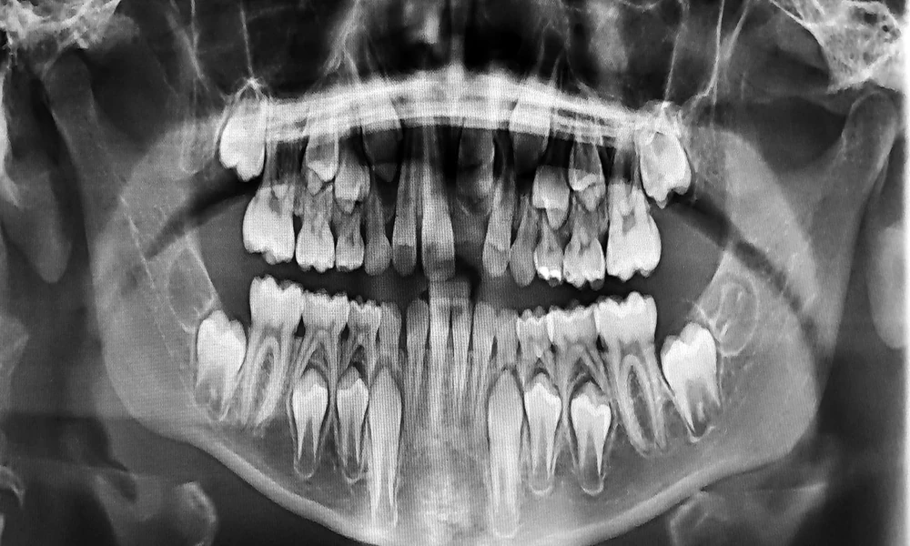
A panoramic X ray helps reveal all the teeth, including those still developing under the gums, missing teeth, extra teeth, root shape, bone levels, and signs of pathology. A cephalometric image, often called a ceph, is different. It is a side view of the head that lets the orthodontist measure how the upper jaw, lower jaw, teeth, and soft tissue relate to one another. If you want a plain language overview of cephalometric radiography, this NCBI resource on cephalometric radiography explains the basics well.
So, what are they actually looking for? First, they study jaw position. Is the upper jaw too far forward, or is the lower jaw set back? Next, they look at tooth inclination. Front teeth can lean too far out or too far in, which affects both the bite and facial support. They also check growth direction. Some patients grow more vertically, which can open the bite, while others grow more horizontally, which changes how the jaws fit together.
What can a cephalometric analysis show that your smile alone cannot?
This is where things get more precise. A cephalometric analysis uses landmarks and measurements to compare your anatomy with expected patterns. That does not mean treatment is based on a single number. It means the orthodontist has a map to guide decisions.
For example, if a child has a developing overbite, the image may show whether the issue is mostly dental, meaning the teeth are positioned poorly, or skeletal, meaning the jaws are growing out of proportion. That difference matters. Dental problems often respond to tooth movement alone. Skeletal problems may call for growth modification, timing changes, or, in adults with severe cases, a discussion about surgical orthodontics.
Research has also looked at how cephalometric methods have changed over time and how different analyses help with diagnosis. This review of cephalometric analyses outlines how these measurements support orthodontic planning. Another study on cephalometric landmarks and interpretation helps show why consistent image reading matters when treatment decisions depend on small anatomical differences.
In practical terms, an orthodontist may be asking questions like these. Are the front teeth flared because of crowding? Is the chin position affecting facial balance? Is there enough room for unerupted teeth? Could airway space or tongue posture be playing a part? This is why what orthodontists look for on X rays and cephalometric images goes far beyond checking whether teeth are crooked.
How do panoramic and cephalometric images compare when planning orthodontic treatment?
When people hear “X ray,” they often assume one image does it all. It does not. Each type serves a different purpose, and together they give the orthodontist a more complete view of your bite and facial structure.
| Image Type | What It Shows Best | Why It Matters in Orthodontics |
|---|---|---|
| Panoramic X ray | All teeth, roots, eruption patterns, missing or extra teeth, general bone levels | Helps detect hidden dental issues and guides space planning |
| Cephalometric image | Jaw relationships, facial profile, tooth angulation, growth direction | Helps diagnose bite problems and plan tooth movement with facial balance in mind |
| Clinical photos and exam | Smile appearance, gum display, bite contact, soft tissue details | Connects the image findings to how your teeth and face look in real life |
That comparison matters because treatment can go off track if one piece is missing. A patient may think the main issue is crowding, while the orthodontic X ray analysis shows a deeper jaw mismatch. Or a parent may worry about a delayed tooth, while the image shows the tooth is blocked by another one and needs a different plan.
What should you do if you want to understand your own orthodontic imaging better?
You do not need to memorize angles or landmark names, but you should feel comfortable asking questions. If the explanation feels rushed or too technical, it is fair to slow it down. This is your face, your bite, and your treatment.
Ask what problem the image confirms. Instead of asking only, “Do I need braces?” ask, “What does this image show about my teeth and jaw?” That question often leads to a clearer answer about whether the issue is spacing, bite, growth, impacted teeth, or facial balance.
Ask how the findings affect treatment choices. If two plans are on the table, such as clear aligners versus braces, or early treatment versus waiting, ask how the X rays and ceph support one option over the other. A good explanation should connect the image to the plan in simple terms.
Ask what needs monitoring over time. Some findings do not call for action right away. A child may need growth watched, or an adult may need root position monitored during treatment. Understanding what the orthodontist is tracking can make the process feel less uncertain.
So where does that leave you if the images seem overwhelming?
It leaves you in a better place than you may think. These images are tools, not judgments. They help an orthodontist spot issues early, explain why a bite works the way it does, and build a plan that fits both function and appearance. Whether you hear the phrase cephalometric images in orthodontics, orthodontic imaging, or simply orthodontist evaluation, the goal is the same. To see what the eye alone cannot see.
If you are moving forward with an orthodontist, ask for a plain language review of your images and what they mean for treatment. A clear explanation can turn a confusing appointment into one that finally makes sense.